Lipid Membrane Nanostructure
Eukaryotic cell membranes (Figure 1) define and enclose the cell are composed of lipids molecules and proteins. The lipid molecules are amphiphilic and self-assemble in an aqueous environment to form a bilayer structure where the hydrophilic headgroups face the aqueous environment and the hydrophobic tailgroups form the interior of the membrane. Associated with the membrane are membrane proteins that perform a wide variety of cellular functions such as transport and signalling.

Figure 1: Schematic representation of a Cell Membrane (Source)
The membrane is sectively permeable, generally less permeable to large, polar solutes and therefore drugs and biomolecules interacting with the cell must first permeate the membrane.
We have used low-noise AFM to study the structure and hydration of gel-phase phopholipid bilayers (Figure 2) and are able to image the bilayer surface with sub-molecular resolution and see that the zwitterionic headgroups of the lipid molecules form ordered arrays of dipoles on the surface. We find that associated with the surface of the bilayer are at least two ordered water layers that present energy barriers to interacting external molecules.
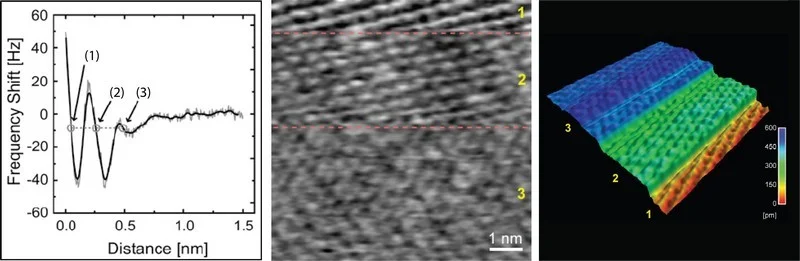
Figure 2: (a) Interaction force between AFM tip and a DPPC bilayer surface showing two oscillatory peaks associated with ordered water layers adjacent to the membrane surface. AFM height image of a DPPC bilayer in PBS buffer in 2D (b) and 3D (c). The surface structure of the adsorbed water layers is seen in regions 2 and 3 and that of the DPPC bilayer in region 3, the lipid headgroup structure and organisation revealed with sub-molecular resolution.
We are currently studying the structure of lipid rafts with sub-molecular resolution using high resolution, low-noise AFM. Lipid rafts are small (sub-100nm) liquid-ordered assemblies of sphingolipids, cholesterol and raft-associated membrane proteins that diffuse within the liquid disordered lipid membrane. Raft and raft-associated membrane proteins have been implicated in a growing number of physiological and pathological cellular processes and hence our interest in them.

Figure 3: Schematic representation of Lipid Raft. A Intracellular space or cytosol. B Extracellular space or vesicle/Golgi apparatus lumen. 1. Non-raft membrane, 2. Lipid raft, 3. Lipid raft associated transmembrane protein, 4. Non-raft membrane protein, 5. Glycosylation modifications (on glycoproteins and glycolipids), 6. GPI-anchored protein, 7. Cholesterol, 8. Glycolipid. (Source)

Figure 4: Phase separation in a DOPC, Sphingomyelin (SM) , and Cholesterol bilayer imaged using dynamic AFM in Sodium Phosphate buffer. The yellow regions show liquid ordered SM/cholesterol bilayer extending about 1nm above the surface of the liquid disordered DOPC/cholesterol (red) bilayer.
References
- Direct Imaging of Lipid-Ion Network Formation under Physiological Conditions by Frequency Modulation Atomic Force Microscopy, Fukuma, T., Higgins, M. J., and Jarvis, S. P., Physical Review Letters, 98, 106101, (2007).
- Direct Imaging of Individual Intrinsic Hydration Layers on Lipid Bilayers at Ångstrom Resolution, Fukuma, T., Higgins, M. J., and Jarvis, S. P., Biophysical Journal, 92, 3603-3609, (2007).
- Lipid Rafts As a Membrane-Organizing Principle, Lingwood, D. and Simons, K., Science, 327, 46 (2009).
- Direct Submolecular Scale Imaging of Mesoscale Molecular Order in Supported Dipalmitoylphosphatidylcholine Bilayers, Sheikh, K. H., Giordani, C., Kilpatrick, J. I., Jarvis S. P., Langmuir, 27, 7, 3749-3753, (2011).
- Direct imaging of salt effects on lipid bilayer ordering at sub-molecular resolution, Ferber, U. M., Kaggwa, G. B., Jarvis, S. P., European Biophysics Journal, 40, 3, 329-338, (2011).