Fluorescence Microscopy
Fluorescence microscopy has become a valuable tool in biological research to visualize and follow processes in cells through the development of fluorescent markers, especially genetically engineered constructs of fluorescent proteins attached to other proteins of interest, so that expression level, cellular distribution and protein interactions can be studied in detail in living cells or organisms. Technological advances such as confocal and total internal reflection microscopy have allowed imaging of very high quality and 3 dimensional sectioning of samples.
The combination of AFM and fluorescence techniques is valuable as both techniques allow different kinds of measurements to be done on the same sample at the same time. AFM can be used to image surfaces at very high, submolecular, resolution as well as to measure forces and mechanical properties and to manipulate cells and molecules mechanically. Fluorescence microscopy can be used simultaneously to image the 3 dimensional structure of cells and to follow a cells reaction to manipulations through its protein expression and distribution.

Figure 1: Cutting of an actin fiber with an AFM cantilever. The fiber then depolymerizes from the cut to where it crosses a different fiber

Figure 2: Cell stained for actin (red), focal adhesions (green) and the nucleus (blue)
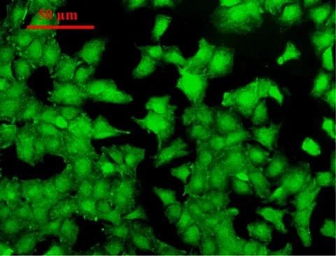
Figure 3: Immunocytochemical investigation of Cyclin E protein expression in osteoblasts. Cyclin E expression is initiated in the middle of G1 phase of the cell cycle and is necessary for progression to S phase.