Biocompatible Materials
One of the main challenges facing the long term success of load-bearing implants and prosthetics, is the lack of predictability. This is largely due to the lack of initial osseointegration and the growth of undesired cell types on the implant surface. The body’s first encounter with an implant is at the cell-surface interface. The colonisation of specific cell types and subsequent tissue development is crucial to the success of the device. Surface properties such as physical topography, chemistry and mechanical properties play a crucial role in the regulation of cellular behaviour such as adhesion, proliferation and differentiation. In order to engineer a biomaterial with optimal surface properties, it is vital to gain an understanding of how such properties affect the response of certain cell types. Currently, nanoscale materials are attracting a lot of attention as a potential biomaterial, due the ability of nanoscale topography to stimulate significant cell responses. Ideally, careful control and modification of the individual mechanical, topographical and chemical surface parameters would encourage the adhesion and proliferation of desired cell types, whilst discouraging adhesion of other types. Such control over surface properties and modifications provides a vital tool in biomaterials engineering.
AFM is a useful tool for the analysis of cell behaviour as live cells can be non-destructively imaged in aqueous physiological conditions, which is a major advantage over other nanoscale imaging techniques. Cellular response is generally evaluated in terms of cell morphology, adherence, size, mobility, proliferation rate and differentiation. AFM can be used to evaluate a large range of these responses, and, the addition of confocal microscopy allows the visualisation of various cellular components, including cytoskeleton and focal adhesions via fluorescent staining. Current work involves the investigation of the response of bone forming osteoblast cells to nanotubular titania surfaces with modified mechanical and chemical properties. In comparison to flat surfaces, cells have been shown to have a higher proliferation rate on all nano-surfaces and exhibit an overall differential response to the nanoscale topography. The differentiation of cells is being investigated using common osteogenic markers, osteocalcin, collagen 1 and bonesialoprotein.
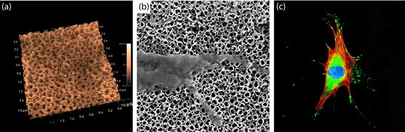
Figure 1: (a) 3D topographical map of the anodised nanotubular titania with an average diameter of 70nm and approximate height (layer thickness) of 500nm. Imaged with a carbon nanotube attached to a conventional AFM tip. (b) Scanning electron micrograph displaying cellular interaction with the nanotubular surface via nanoscale filopodial extensions. Cell filipodia are believed to be a sensory tool used by cells to investigate the surrounding environment. Image dimensions 3.2 x 3.2 um (c) Confocal image of a cell on nanoscale titanium stained for nucleus (blue), cytoskeleton (red) and focal adhesions (green). The confocal image provides qualitative and quantitative information for comparison of cells on various modified surfaces. Image dimensions 190 x 190um.
References
A collagen-glycosaminoglycan scaffold supports adult rat mesenchymal stem cell differentiation along osteogenic and chondrogenic routes, Farrell, E., O'Brien, F., Doyle, P., Fischer, J., Yannas, I., Harley, B., O'Connell, B., Prendergast, P., and Campbell, V., Tissue Engineering, 12, 459-468, (2006).